Friday 30 August 2013
Sarah Hillary
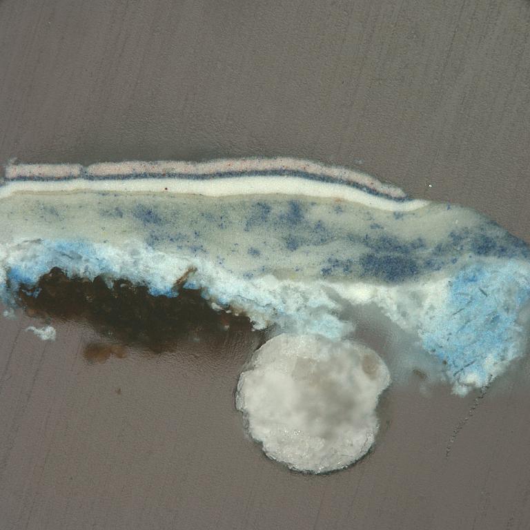
A tiny sample of paint embedded in resin can be made into a cross-section. They allow us to see the layering structure of the painting down to a microscopic level and we can analyse the individual layers as a consequence.

A cross-section from the painting Cross 1959 by Colin McCahon
I have been making cross-sections for many years but wanted to improve my technique and Getty Conservation Institute (GCI) Scientist, Alan Phenix, agreed to show me the finer details of his method. Alan’s work at the GCI focuses on paint analysis, primarily to assist Getty Museum painting conservators with the paintings in their care.
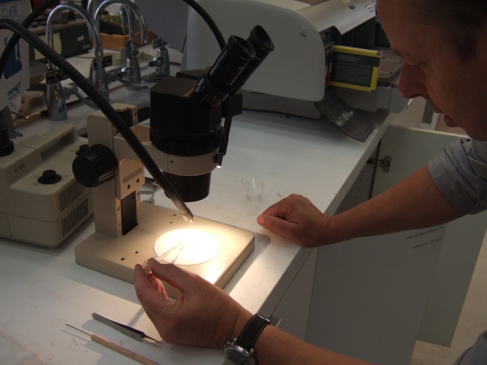
Sampling from paintings is only done if absolutely necessary and great care is taken in finding a suitable location. The cross-section sample will be smaller than a pin-head and from an existing damage. Alan looks through the microscope to place the edge of the cross-section on a glass slide where it is secured. A plastic mould is placed around it, resin poured in and label inserted to the side.

GCI Scientist, Alan Phenix, pouring resin into the mould to prepare a cross-section.
The resin is placed in a chamber and cured by exposure from ultra-violet (UV) radiation for 25 minutes. UV setting resins are also commonly used by dentists today. Now the cross-section is ready for sanding and polishing followed by microscopic examination and digital photography.

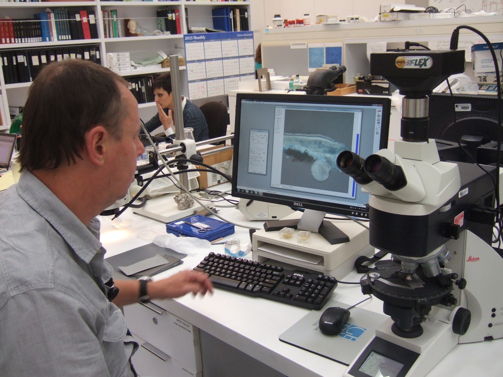
Alan looking at the McCahon cross-section
In the past the process of making a cross-section would have taken several days, where today we can get much better results in a couple of hours. Alan helped me to improve the surface of a cross-section from the McCahon painting Cross 1959 and he took a photo. Pity about the air bubble in the resin next to the sample, but I won’t make that mistake again!
The Getty will be publishing their cross-section technique online in the near future and more information about Alan can be found here:
http://www.getty.edu/conservation/publications_resources/newsletters/22_3/gcinews9.html